(Figs 1-15)
Patina oussagari Therond, 1975: 749 [Tanzania].
Theropatina oussagarai : Mazur, 1984b: 255.
Redescription (Mazur & Ohara, 2000b).
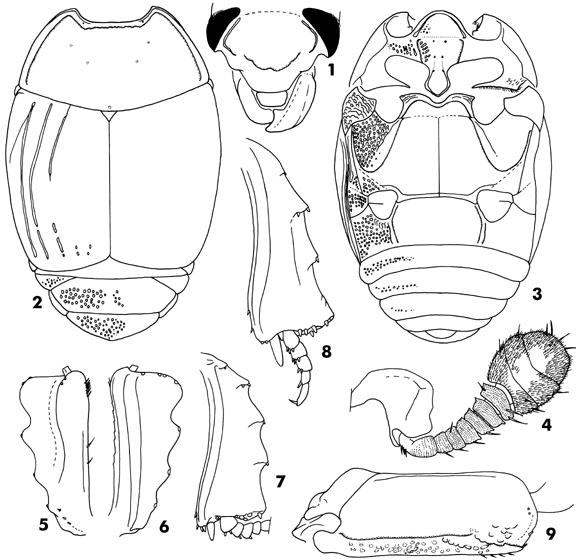
Body (Fig. 2) oval, black; funicule of antennae and tarsi dark brown. Body length (male, type, in mm), PPL, 8.8; PEL, 6.8; APW,2.0; PPW, 5.0; PL, 2.4; EL, 3.8; EW, 5.6; ProL, 1.1; ProW, 3.4; PyL, 1.4; PTL, 1.8; MSTL, 1.7; MTTL, 2.0.
Antennal club clear suture (Fig. 4), the suture almost straight and not interrupted medially. Ratio in width of pronotum to head 2.94. Front of head feebly depressed; surface sparsely and finely punctate, the punctures separated by about three times their diameters. Frontal stria of head (Fig. 1) carinate and interrupted at lateral one-fourth. Labrum transverse oblong and its surface flat, the anterior margin round. Surface of mandible without carina.
Pronotum (Fig. 2) feebly convex medially; marginal stria completely impressed laterally and broadly interrupted behind head; outer lateral pronotal stria well impressed, carinate and complete laterally and anteriorly, the medio-apical portion behind head almost straight and storngly crenate. Surface of pronotum sparsely clothed with fine punctures; two pairs of slight impression present on medio-anterior area. Antescutellar area with a slight impressed point.
Epipleura with two marginal striae, the outer stria strongly carinate. Elytral marginal stria complete and strongly carinate. External subhumeral stria slightly impressed on basal two-thirds. Internal subhumeral stria absent. Oblique humeral stria slightly impressed on basal two-fifths. First to 2nd dorsal striae complete; 3rd present on basal half and some rudiments on apical fourth; 4th and 5th striae only represented by few rudiments apically; sutural stria absent. Surface of elytra shining and impunctate.
Propygidium densely with large and longitudinal oblong punctures, the punctures separated by half their diameter; interspace among the large punctures and broad area along margin clothed with fine punctures; surface with feebly depression on each lateral side. Pygidium flat and densely covered with large, round and deep punctures which are separated by half to twice their diameter.
Prosternal lobe (Fig. 3) convex medially, its anterior margin round; marginal stria complete and carinate, the lateral end with deep fovea; secondary marginal stria absent; surface sparsely and coarsely punctate. Prosternal process with carinal striae on posterior half, the striae united each other on posterior area and impressed completely along posterior margin; surface impunctate; posterior margin cuspid outwardly, but apex round. One lateral prosternal stria present and strongly carinate.
Mesosternum transverse; surface clothed with fine and shallow punctures along marginal stria; anterior margin deeply emarginate medially; marginal stria of mesosternum completely impressed, carinate and sinuate medially; other short striae well impressed behind each antero-lateral angle. Meso-metasternal suture slightly arcuate anteriorly. Intercoxal disk of metasternum flat and impunctate. Lateral metasternal stria carinate, extending obliquely and posteriorly, then, strong curved outwardly, the apical end attained at antero-lateral corner. Post-mesocoxal stria straight and impressed on basal half. Lateral metasternal disk covered with large, semicircular and shallow punctures which are sometimes fused with each others.
Intercoxal disk of 1st abdominal sternum impunctate; two lateral striae present on each side, the inner one complete and carinate, the outer one present on posterior half. Lateral disk irregularly and densely covered with coarse punctures and longitudinal rugae.
Protibia (Fig. 5, 6) with 4 spines on outer margin (not including a spine at apical outer angle), their bases denticulate, and a pair of spines on inner angle and 4 or 5 spine present on apical margin (but it seems that the state of the only type specimen available is imcomlete, with dermal structure and spines defaced to some extent) . Mesotibia (Fig. 7) with 3 spines on outer margin, and 9 spines present on apical margin; ventral surface with a row consisting of 2 or 3 spines. Metatibia (Fig. 8) with 3 dental spines on outer margin, and 10 spines present on apical margin; ventral surface with two small spines. Ventral surface of profemur (Fig. 9) with transverse stria, and clothed with coarse punctures along posterior margin.
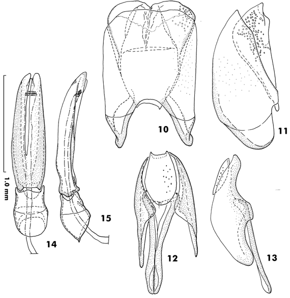
Male genitalia (Fig. 10-15). Posterior margin of eighth sternite straight, not divided. Ninth tergite well sclerotized and with antero-lateral projections, but not like stick. Spicule is Y-letter shaped. Tenth tergite oblong, the posterior margin broadly emarginate. Ratio in length of parameres to basal piece about 2.64. Lateral sides of parameres paralleled on anterior half, thence convergent apically on posterior half; parameres not fused on apical two-thirds of dorsal surface. Median lobe simple, probably extruded from the dorsal side of tegmen.
Specimen examined. TANZANIA. Holotype, male, Holotype (red label), Tanzanie: Mts Uluguru, Kiroka, for héliophile, alt, 725 m, 27-31-V-(19)71, Coll. Mus. Tervuren, Mission Mts Uluguru, L. Berger, N. Leleup, J. Debecker, V/VIII/71, Patina oussagarai nov. sp. J. Therond det., 1924 (MTF: #MO-00-145).
Emmamushi.Home/Histerinae/Platysomatini/Theropatina/References