Plaesius (Plaesius) javanus Erichson, 1834: 102.
Redescription (Ohara & Mazur, 2000).
Body oblong oval, depressed medially, black and shining; antennal club, maxillary palpi and setae of legs reddish brown. Biometric data are given in Table 1.
Antennal club (Fig. 12B) with U-shaped sutures, which are complete and distinct. Ratio in width of pronotum to head about 2.08. Front of head (Fig. 12A) flat; surface densely covered with fine punctures that are separated by their own diameter or twice the diameter. Frontal stria of head shortly interrupted at middle and sinuate at lateral angle. Supraorbital stria absent. Labrum transverse and short, the anterior margin sinuate at middle. One denticle present on inner margin of mandible.
Pronotum (Fig. 12C) feebly depressed medially; marginal stria complete laterally and shortly interrupted behind head; outer lateral pronotal stria complete and well impressed, its outer edge cariniform. Surface of pronotum with coriaceous ground sculpture and evenly covered with microscopic punctures that are separated by two to five times their diameter and are sparsely intermingled with fine punctures; inside area of outer lateral stria coarsely and densely punctate, the punctures becoming denser behind apical angle; coarse punctures present along basal margin on lateral fourth. Antescutellar area with a longitudinal impression.
Epipleura with two epipleural marginal striae; the inner (near epipleural margin) stria present on apical half and the outer complete and smoothly curved inwardly at middle. Elytral marginal stria (Fig. 12C, D) complete on epipleura, its inner edge cariniform; the apical end of the stria extended inwardly along posterior margin of elytron and connected with the apical end of sutural stria at posterior inner corner of elytron. External subhumeral stria deeply impressed and sinuate on basal third. Internal subhumeral stria present on apical third, the apical portion curved inwardly. Oblique humeral stria slightly impressed on basal third. First dorsal elytral stria complete and deeply impressed; 2nd stria present on apical half; 3rd stria on apical fourth; 4th stria shortly on apical eighth; 5th stria absent; sutural stria present on apical half. Surface of elytra with coriaceous ground sculpture and sparsely covered with microscopic punctures; a transverse narrow band of coarse and dense punctures along inside of elytral posterior marginal stria,the punctures are separated by about their own diameter.
Propygidium (Fig. 12C) densely covered with longitudinal large and shallow punctures that are separated by about half their diameter, the punctures becoming smaller laterally and posteriorly. Pygidium densely covered with large, round and deep punctures that are separated by one-third to half their diameter.
Prosternal lobe (Fig. 12D) broad and feebly convex medially, its anterior margin arcuate outwardly; marginal stria completely incised, its outer edge subcariniform; surface irregularly and coarsely punctate. One lateral prosternal stria present, the outer edge of striae strongly carinate. Prosternal process flat and finely punctate, with two distinct carinal striae on anterior two-thirds that are parallel on apical half and divergent on posterior half; posterior margin round.
Mesosternum (Fig. 12D) transverse and flat; surface evenly and finely punctate, the punctures separated by three to five times their diameter; anterior margin deeply emarginate at middle; marginal stria of mesosternum broadly interrupted medially and abbreviated laterally on posterior fourth, its outer edge subcariniform; another short stria present behind antero-lateral angle. Meso-metasternal suture finely impressed. Intercoxal disc of metasternum evenly covered with fine punctures. Lateral metasternal stria extending posteriorly, its outer edge cariniform, the apical end attaining nearly to posterior two-third of metasternal-metepisternal suture. Post-mesocoxal stria absent. Lateral metasternal disc covered with transverse rugae, the anterior edges of rugae subcariniform; area at posterior three-fourths of lateral disc projected posteriorly.
Intercoxal disc of 1st abdominal sternum (Fig. 12D) with punctation similar to that of intercoxal disc of metasternum, the area before posterior corner coarsely punctate; one lateral stria present on each side, shortened on posterior sixth, its outer edge subcariniform, the striae arcuate and convergent anteriorly. Lateral disc densely covered with longitudinal rugae.
Protibia (Fig. 12E, F) with two large denticles on outer margin; dorsal surface with a sinuate and deep tarsal groove; a longitudinal stria incised along the groove and also along inner margin; on ventral side, about 50 to 55 short robust spines present on apico-lateral margin; ventral surface densely and coarsely punctate and striate along inner margin, the outer edge of the stria cariniform. Mesocoxa without carina. Meso- and metatibiae (Fig. 12G, H) with about 65 robust spines on outer margin, the spines represented by 3 longitudinal rows. Ventral surface of profemur with transverse rugae on posterior half.
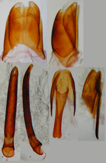
Male genitalia (Fig. 13). Eighth sternite divided into two lobes, which are not prolonged apically. Ninth tergite without stick-like antero-lateral projections. Spicule Y-shaped. Ratio in length of parameres to basal piece about 4.2; basal piece short. Lateral sides of parameres parallel, then strongly convergent on apical eighth; parameres fused on dorsal surface, but separated on apical eighth. Median lobe simple.
Female genitalia (Fig. 12I, J). Spermatheca weakly sclerotized and round.
Specimens examined. Thailand. 1 female, Cheng Dao, 8-IV-1981, M. Ito. Malaysia. Proper (Peninsula): 1 female, Tapah, Perak, 16-III-1999, T. Yoshida; 1 female, Ditto, 8-VIII-1978, K. Sugiyama. Indonesia. Borneo: 3 males, 3 female, Mt. Bawang, alt. 250300 m, western Kalimantan, VIII, X-1990, VII-1991. Sumatra: 1 male, Air Mancur, Nr. Bukit Tinggi, I-1989, Jamoan. Celebes: 1 female, Palolo Palu, IX-1995. All specimens are deposited in SEHU.
Distribution. Oriental Region.
Fig. 12. Plaesius (Plaesius) javanus. A: Head. B: Antenna. C: Adult, dorsal view. D: Ditto, ventral view. E: Left protibia, dorsal view. F: Ditto, ventral view. G: Left mesotibia, ventral view. H: Left metatibia, ventral view. I: Female genitalia, ventral view. J: Spermatheca. (Ohara & Mazur, 2000).
Fig. 13. Plaesius (Plaesius) javanus. A: Aedeagus, dorsal view. B: Ditto, lateral view. C: 9th and 10th tergites and spicule, dorsal view. D: Ditto, lateral view. E: 8th tergite and sternum, dorsal view. F: Ditto, lateral view. (Ohara & Mazur, 2000).
MO-02-011P (Malaysia: Borneo, Sabah).