Placodes senegalensis (Paykull, 1811)
Hister senegalensis Paykull, 1811: 13 [Guinea].
Placodes senegalensis: Marseul, 1853: 232: Ohara & Mazur, 2000.
Redescription (Ohara & Mazur, 2000).
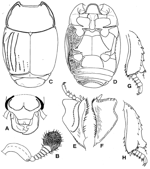
Body oblong-oval, brownish black and shining; maxillary palpi, club of antenna reddish brown. Body length: PPL 9.8; PEL 8.6; APW 2.5; PPW 5.8; PL 3.2; EL 4.9; EW 6.4; ProL 1.6; PROW 3.5; PyL 1.7; PTL 2.0; MSTL 2.2; MTTL 2.5.
Antennal club (Fig. 10B) with V-shaped sutures. Ratio in width of pronotum to head 2.63. Front of head (Fig. 10A) flat. Supraorbital stria deeply impressed laterally; frontal stria complete and cuspidate inwardly at middle, its outer edge well cariniform. Disc of head evenly covered with fine punctures that are separated by about four times their diameter. Labrum transverse, feebly depressed medially, the anterior margin almost straight. Mandible elevated laterally and excavate at base; one denticle present on inner margin of mandible.
Pronotum (Fig. 10C) feebly convex; the anterior margin broadly and regularly emarginate; marginal stria completely impressed laterally and anteriorly, on the anterior portion the stria being a little at distance from margin; outer lateral stria broad, deeply impressed and complete laterally, and a little sinuate at middle, the apical end extended inwardly and shortly; surface covered with fine punctures that are separated by about three times their diameter. Antescutellar area with a short longitudinal impression.
Epipleura with two marginal striae, the outer one complete and the inner one present on apical half. Elytral marginal stria complete, extending inwardly along apical margin, thence curved anteriorly at middle corner of elytron, the outer edge subcariniform. Outer subhumeral stria (Fig. 10C) present on basal half. Inner subhumeral stria present on apical two-thirds. Oblique humeral stria slightly impressed on basal third. First to 3rd dorsal striae complete, the basal end of 1st stria bent inwardly and 3rd stria strongly sinuate; 4th and 5th dorsal striae present on apical one-third, but the basal half of the striae represented by a row of coarse punctures; the sutural stria consisted of several coarse punctures. Surface of elytra evenly covered with fine punctures that are separated by about twice their diameter; a narrow band of coarse punctures along the posterior margin.
Propygidium transverse and elevated behind at lateral corner; disc densely covered with large punctures that are separated by about their own diameter; interspace among the large punctures covered with alutaceous ground sculptures. Pygidium feebly depressed on apical half; disc densely covered with large punctures which are separated by one-third to half their diameter, the large punctures becoming smaller apically.
Prosternal lobe (Fig. 10D) convex medially; anterior margin round; marginal stria complete and its outer edge subcariniform; secondary stria shortly impressed basally; surface of lobe shining and sparsely and finely punctate. Prosternal process feebly elevated, without carinal stria; posterior margin round. One lateral prosternal stria present, the outer edge cariniform.
Anterior margin of mesosternum (Fig. 10D) deeply emarginate; marginal stria interrupted medially; two secondary striae present behind each antero-lateral angle. Meso-metasternal suture slightly impressed and angulate at middle. Disc of mesosternum sparsely and finely punctate, the punctures separated by five to ten times their diameter. Lateral metasternal stria extending obliquely, the outer edge subcariniform and the apical end attaining to apical two-thirds of lateral disc. Post-mesocoxal stria absent. Intercoxal disc of metasternum smooth, covered with fine punctures that are separated by three to five times their diameter. Lateral disc densely covered with irregular, transverse and carinate rugae; strong angled elevation present on postero-lateral area, the inner end of the elevation placed in front of inner corner of metacoxa and the outer end attaining to middle of metasternal-metepisternal suture.
Intercoxal disc of 1st abdominal sternum (Fig. 10D) smooth and finely punctate, the punctures becoming coarser along lateral stria and posterior margin; lateral stria complete. Lateral disc densely covered with longitudinal rugae. Lateral side of second and fifth sterna densely covered with large punctures.
Protibia (Fig. 10E) with 1 large denticle on outer margin, and a large spines on inner angle and 12 spines present on apical margin (including a spine at apical outer angle). Mesocoxa without distinct carina. Mesotibia (Fig.10G) with 5 spines on outer margin, and 17 spines on apical margin, of which 6 spines are compressed together on the outer tooth (expanded angle); ventral surface with a spiny row represented by 4 spines along the outer margin. Metatibia (Fig. 10H) with 7 spines on outer margin, and with 15 spines on apical margin, of which 4 spines are compressed together on the outer tooth (expanded angle); ventral surface with a spiny row represented by 4 spines along the outer margin. Ventral surface of profemur densely covered with transverse oblong punctures.
Male genitalia (Fig. 11). Eighth sternite divided into two lobe. Ninth tergite without antero-lateral projections stick-like in shape. Spicule Y-shaped. Ratio in length of parameres to basal piece about 7.1; basal piece short. Lateral sides of parameres parallel, the apex truncate; parameres fused on dorsal surface but separated on apical fifth. Median lobe simple.
Specimen examined. Congo. 1 male, Belg. Congo, ex. coll. F. Kessel, Mus. Zool. Polonicum Warszawa, 19/46, Placodes senegalensis Det. S. Mazur (MIZPAN).
Distribution. Guinea, tropical Africa.
Fig. 10. Placodes senegalensis. A: Head. B: Antenna. C: Adult, dorsal view. D: Ditto, ventral view. E: Left protibia, dorsal view. F: Ditto, ventral view. G: Left mesotibia, ventral view. H: Left metatibia, ventral view. (Ohara & Mazur, 2000).
Fig. 11. Placodes senegalensis. A: Aedeagus, dorsal view. B: Ditto, lateral view. C: Apex of aedeagus, ventral view. D: 9th and 10th tergites and spiclue, dorsal view. E: Ditto, lateral view. F: 8th tergite and sternum, dorsal view. G: Ditto, lateral view. (Ohara & Mazur, 2000).