Idister (Diister) platysomoides Mazur, 1989: 736.
Redescription. (Ohara & Mazur, 2000).
Body oblong-oval, brownish black and shining; tibiae, tarsi, maxillary palpi, funicle of antenna reddish brown. Body length in mm: PPL 6.2; PEL 5.2; APW 1.55; PPW 3.35; PL 1.9; EL 3.05; EW 3.6; ProL 0.45; PROW 1.95; PyL 0.9; PTL 1.2; MSTL 1.25; MTTL 1.45.
Antennal club (Fig. 8B) with V-letter shaped sutures, the suture between 9th and 10th interrupted medially. Ratio in width of pronotum to head 2.68. Front of head (Fig. 8A) flat. Supraorbital stria impressed laterally and frontal stria sinuate and finely incised medio-anteriorly but interrupted at middle. Disc of head evenly covered with microscopic punctures which are separated by about three times their diameter. Labrum transverse, the anterior margin slightly emarginate medially. One denticle present on inner margin of mandible.
Pronotum (Fig. 8C) feebly convex; the anterior margin broadly and regularly emarginate; marginal stria completely impressed on lateral margin and interrupted on median third of anterior portion; outer lateral stria complete laterally, and inwardly extending apically, then united with marginal stria; surface covered with coriaceous microsculpture and sparsely and finely punctate. Antescutellar area with a short longitudinal impression.
Epipleura (Fig. 8D) with complete epipleural marginal and elytral marginal striae, their outer edges subcariniform. Subhumeral striae (Fig. 8C) absent. Oblique humeral stria slightly impressed on basal third. First dorsal stria complete; 2nd shortened on basal tenth to one-third (holotype), usually the basal half of the stria finely incised; 3rd absent (holotype) or represented by fragmented impression on apical sixth, and finely incised on basal third; 4th, 5th dorsal and sutural striae absent. Surface of elytra evenly covered with fine punctures that are separated by about four times their diameter.
Propygidium (Fig. 8C) smooth, sparsely and finely punctate, the punctures separated by about four times their diameter, and covered with alutaceous microscopic punctures; only one coarse puncture present on each latero-median area. Pygidium densely covered with large and round punctures that are separated by one-third to half their diameter, the large punctures absent along latero-apical margin.
Marginal stria of prosternal lobe (Fig. 8D) present on apical half; secondary stria impressed along lateral margin on basal half; surface of lobe densely covered with fine punctures which are separated by twice to six times their diameter. Prosternal process without carinal stria; posterior margin round. Two lateral prosternal striae present.
Anterior margin of mesosternum (Fig. 8D) emarginate; marginal stria complete, the outer edge subcariniform. Meso-metasternal suture slightly impressed and angulate at middle; surface finely punctate, the punctures separated by about twice their diameter. Lateral metasternal stria extending obliquely, the outer edge subcariniform and the apical end attaining to lateral sixth. Post-mesocoxal stria absent. Intercoxal disc of metasternum smooth, with punctation similar to that of mesosternum. Lateral disc irregularly with several large, round and shallow punctures, sometimes the punctures fused with each other to form transverse rugae.
Intercoxal disc of 1st abdominal sternum (Fig. 8D) with punctation similar to that of metasternum; lateral stria complete. Lateral disc densely covered with longitudinal rugae. Lateral side of second and third sterna with transverse stria.
Protibia (Fig. 8E, F) with 4 spines on outer margin (not including a spine at apical outer angle), their bases strongly denticulate, and with a pair of spines on inner angle and 2 spines present on apical margin (including a spine at apical outer angle). Mesocoxa with distinct carina. Mesotibia (Fig. 8G) with 5 dental spines on outer margin, and 7 spines present on apical margin; ventral surface with a spiny row represented by two spines basally. Metatibia (Fig. 8H) with 3 spines on outer margin, and 6 spines present on apical margin; ventral surface without spiny row but with a distinct carina. Ventral surface of profemur covered with transverse rugae only on posterior fourth.
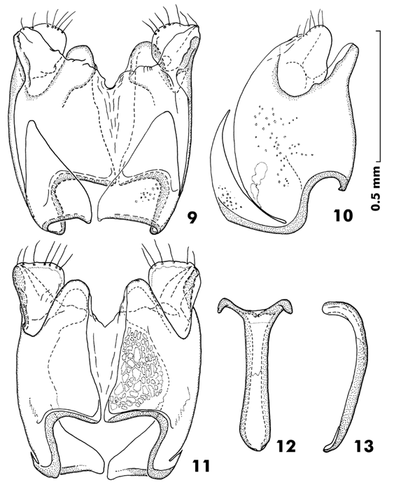
Male genitalia (Fig. 9). Eighth sternite divided into two lobe. Ninth tergite with antero-lateral projections stick-like in shape. Spicule simple, elongate. Ratio in length of parameres to basal piece about 1.19; basal piece long. Lateral sides of parameres parallel, the apex truncate; parameres fused on dorsal surface. Median lobe simple.
Specimens examined. Papua New Guinea. Male (Paratype), Mt. Hagen, Rokina Bayer Valley, 21-IV-1979, W.G. Ullrich (SMC).
Distribution. Papua New Guinea.

Fig. 8. Idister (Diister) platysomoides. A: Head. B: Antenna. C: Adult,
dorsal view. D: Ditto, ventral view. E: Left protibia, dorsal view. F: Ditto,
ventral view. G: Left mesotibia, ventral view. H: Left metatibia, ventral
view. (Ohara & Mazur, 2000).
Fig. 9. Idister (Diister) platysomoides. A: Aedeagus, dorsal view. B: Ditto, lateral view. C: 8th tergite and sternum, dorsal view. D: Ditto, lateral view. E: 9th and 10th tergites, dorsal view. F: Ditto, lateral view. G: Spicule, dorsal view. H: Ditto, lateral view. (Ohara & Mazur, 2000).