Idister (Diister) omalodellus Mazur, 1989: 735.
Redescription. (Ohara & Mazur, 2000).
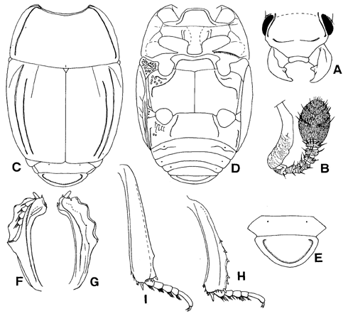
Body oblong oval, slightly convex medially, black and shining; antenna, mouth parts and legs reddish brown. Body length; PPL, 5.30; PEL, 4.65; APW, 1.37; PPW, 3.01; PL, 1.62; EL, 2.65; EW, 3.28; ProL, 0.54; ProW, 1.76; PyL, 0.83; PTL, 1.32; MSTL, 1.37; MTTL, 1.72.
Antennal club (Fig. 6B) with V-shaped sutures, which are interrupted medially. Ratio in width of pronotum to head 2.46. Front of head feebly depressed medially; surface sparsely covered with fine punctures that are separated by about three times their diameter. Frontal stria of head well impressed but interrupted at middle and lateral angle, its outer edge subcariniform. Supraorbital stria absent. Labrum transverse and short, the anterior margin emarginate inwardly. One denticle present on inner margin of mandible.
Pronotum (Fig. 6C) feebly convex post-medially; marginal stria complete laterally and anteriorly; outer lateral pronotal stria completely impressed, its outer edge cariniform. Surface of pronotum irregularly and densely covered with fine punctures that are separated by two to five times their diameter. Antescutellar area with a slight longitudinal impression.
Epipleura with marginal stria on apical half, its inner (near epipleural margin) edge subcariniform. Elytral marginal stria (Fig. 6D) complete and sinuate at middle, its inner edge subcariniform. Subhumeral stria absent. Oblique humeral stria (Fig. 6C) slightly impressed on basal third. First dorsal elytral stria completely impressed, its outer edge cariniform; 2nd stria shortened on basal sixth; 3rd stria slightly impressed on basal third; 4th, 5th and sutural striae absent. Surface of elytra evenly covered with fine punctures that are separated by about four times their diameter.
Propygidium (Fig. 6C, E) sparsely covered with fine punctures that are separated by about four times their diameter and with one coarse punctures present at each antero-lateral half. Pygidium (Fig. 6E) striate along posterior margin, the outer edge of the stria strongly carinate; area between the stria and margin of pygidium elevated; surface evenly and sparsely covered with fine punctures that are separated by four times their diameter.
Prosternal lobe (Fig. 6D) broad and feebly convex medio-anteriorly, its anterior margin truncate; marginal stria complete; surface sparsely covered with fine punctures that are separated by about ten times their diameter. Prosternal process flat and finely punctate sparsely, without carinal striae; posterior margin round. Two lateral prosternal striae present and their outer edges strongly carinate.
Mesosternum (Fig. 6D) transverse and flat; surface sparsely and finely punctate, the punctures separated by three to ten times their diameter; anterior margin feebly emarginate medially; marginal stria of mesosternum complete, its outer edge cariniform. Meso-metasternal suture finely impressed and angulated at middle. Intercoxal disc of metasternum with similar punctation of intercoxal disc of mesosternum. Lateral metasternal stria extending posteriorly, curved outwardly and then strongly curved posteriorly near metasternal-metepisternal suture, its outer edge cariniform, the apical end attaining nearly to middle of metasternal-metepisternal suture. Post-mesocoxal stria present along posterior margin of mesocoxa. Lateral metasternal disc densely covered with large, round and shallow punctures.
Intercoxal disc of 1st abdominal sternum (Fig. 6D) sparsely covered with fine punctures; lateral stria present on each side and shortened on apical eighth, its outer edge subcariniform. Lateral disc densely covered with large longitudinal oblong punctures on inner anterior half; one longitudinal stria present along lateral margin; one transverse stria present on each lateral third of 2nd abdominal sternum.
Inner margin of protibia (Fig. 6F, G) strongly curved inwardly on apical fifth; 5 denticles present on outer margin; dorsal surface with rather straight tarsal groove. Mesocoxa with short carina on anterior half. Mesotibia (Fig. 6H) dentate with 5 spines on outer margin and 8 spines on apical margin; ventral surface without spiny row. Metatibiae (Fig. 6I) with 1 spine on outer margin and 7 spines on apical margin; ventral surface without spiny row. Ventral surface of profemur without stria on posterior margin.
Male genitalia (Fig. 7). Eighth sternum divided into two lobe, the lobe elongate posteriorly and curved inwardly at apex. Ninth tergite sclerotized and with antero-lateral projection stick-like in shape. Spicules oblong. Ratio in length of parameres to basal piece about 2.7; basal piece rather short. Lateral side of parameres parallel, the apex expanded laterally and angulate laterally; parameres fused on dorsal surface, but separated on apical third. Median lobe simple.
Specimen examined. Papua New Guinea. 1 male, Morobe, Ung. Gurakor, XII-1979, W.G. Ullrich (SMC).
Distribution. Papua New Guinea.
Fig. 6. Idister (Diister) omalodellus. A: Head. B: Antenna. C: Adult, dorsal view. D: Ditto, ventral view. E: Propygidium and pygidium. F: Left protibia, dorsal view. G: Ditto, ventral view. H: Left mesotibia, ventral view. I: Left metatibia, ventral view. (Ohara & Mazur, 2000).
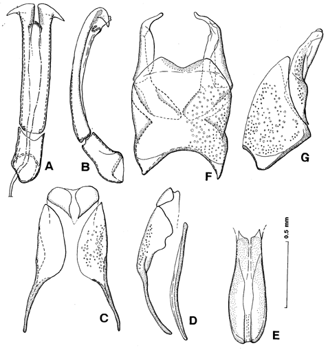
Fig. 7. Idister (Diister) omalodellus. A: Aedeagus, dorsal view. B: Ditto,
lateral view. C: 9th and 10th tergites, dorsal view. D: ditto and spicule,
lateral view. E: Spicule, dorsal view. F: 8th tergite and sternum, dorsal
view. G: Ditto, lateral view. (Ohara & Mazur, 2000).