Atholus philippinensis (Marseul, 1854)
Hister philippinensis Marseul, 1854, 547.
Hister philippensis (sic): Gemminger et Harold, 1868, 771.
Hister (Atholus) philippinensis: Bickhardt, 1913, 173 [Hoozan, Taihorin]; Miwa, 1931, 57 [Hoozan, Taihorin]; Kamiya and Takagi, 1938, 31.
Hister sectator Lewis, 1901, 375, synonymized by Bickhardt, 1917, 194.
Atholus sectator: Lewis, 1906, 402.
Atholus philippinensis: Lewis, 1906, 402; 1915, 55; Ohara, 1999: 32.
Description on the basis of the Taiwanese material. (Ohara, 1999b: Figs. 19-21)
Female. Biometric data as follows (in mm): PPL 4.65, PEL 4.3, APW 1.4, PPW 3.0, PL 1.5, EL 2.0, EW 3.35, ProW 2.0, ProL 0.7, PyL 0.9, PTL 0.95, MSTL 0.85, MTTL 1.1. Body oval, feebly depressed, black and shining; tibiae, tarsi, antennae and mouth parts dark brown.
Frontal stria of head (Fig. 19A) round, complete and deeply impressed. Disk impunctate, wholly clothed with coriaceous microsculpture. Labrum transversely oblong.
Pronotal sides arcuate and strongly convergent apically. Apical angle acute. Marginal stria laterally complete and broadly interrupted behind head. Lateral pronotal stria (Fig. 19B) deeply impressed, sparsely crenate and complete, the lateral portion rather distant from the margin and its basal end reaching to basal fourth of pronotal length. Disk of pronotum without punctation, wholly clothed with coriaceous microsculpture; the narrow posterior band represented by coarse punctures. Antiscutellar area with a short longitudinal puncture.
Marginal epipleural stria present on apical half. Elytral marginal stria complete and carinate. External subhumeral stria (Fig. 19B) abbreviated on basal one-eighth and apical one-sixth. Internal subhumeral stria absent. Oblique humeral stria lightly impressed on basal third. First to 3rd dorsal striae complete, and densely and coarsely crenate. Fourth dorsal stria present on apical half. Fifth and sutural striae present on apical third. Disk evenly and sparsely covered with fine punctures, which are separated by about four times their diameter; the mediobasal area clothed with coriaceous ground sculpture .
Propygidium (Fig. 20C) densely covered with large, round and shallow punctures, which are separated by one to three times their diameter; interspace among the large punctures irregularly and sparsely covered with fine punctures, which are separated by two to five times their diameter. Pygidium densely and coarsely punctate, the punctures separated by about their own diameter to half the diameter and becoming sparser apically; interspace among the coarse punctures densely clothed with fine punctures. Propygidium and pygidium with alutaceous ground sculpture.
Anterior margin of prosternal lobe (Fig. 20E) round; marginal stria deeply impressed, carinate and shortly interrupted at middle; disk coarsely punctate, the punctures separated by one to three times their diameter. Prosternal keel narrow, the anterior half descending; carinal stria absent; lateral disk coarsely punctate. Lateral prosternal stria deeply impressed, carinate and complete.
Anterior margin of mesosternum (Fig. 20E) outwardly arcuate; marginal stria clearly impressed and complete; another stria present behind each anterolateral angle; disk sparsely covered with fine punctures. Meso-metasternal suture complete, angulate at middle. Lateral stria of metasternum (Fig. 20F) deeply impressed, carinate, extending obliquely and posteriorly, beginning from lateral fourth of meso-metasternal suture, and united with the oblique stria that extends inwardly from the middle of metasterno-metepisternal suture; post-mesocoxal stria extending posteriorly and strongly curved along the posterior margin of mesocoxa, and attaining to the middle of metasterno-mesepimeral suture; punctation of intercoxal disk of metasternum similar to that of mesosternum; lateral disk of metasternum densely covered with large and round punctures, which are separated by about half their diameter and become smaller inwardly, and interspace among the large punctures with alutaceous ground sculpture.
Punctation of intercoxal disk of 1st abdominal sternum similar to that of metasternum; lateral stria complete.
Protibia (Fig. 19D, E) with 4 denticles on outer margin and 5 small denticles on apical margin; ventral surface with 5 small denticles along outer margin. Profemoral stria deeply impressed and complete.
Male genitalia as shown in Fig. 21 on the basis of the specimen form western Kalimantan, Indonesia.
Female genitalia as shown in Fig. 19F.
Specimens examined.
Taiwan. [Proper] Nantou Hsien: Nanshanchi, Puli (1 female, 3/iv/1986), M. Ohara.
Indonesia. [Kalimantan] Mt. Bawan, alt. 250300 m, western Kalimantan, (1 male, x/1990), native collector.
Distribution.
Taiwan; Philippines; Malaysia; Burma; Vietnam; Borneo; Java; Sumatra; India; southern China.
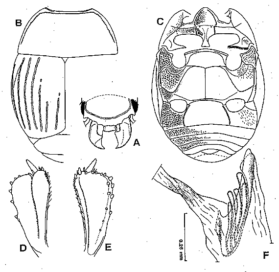
Fig. 19. Atholus philippinensis (Marseul). A: Head, frontal view. B: Pronotum and left elytron. C: Ventral side of adult. D: Left protibia, dorsal view. E: Ditto, ventral view. F: Female genitalia, spermatheca, vagina and bursa copulatrix, lateral view (left side). [no. 9930, Nanshanchi]. (Ohara, 1999b).
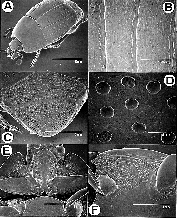
Fig. 20. Atholus philippinensis (Marseul). A: Adult, female, oblique view. B: Surface of elytron, dorsal view. C: Propygidium and pygidium, caudal view. D: Punctation of pygidium. E: Prosternum, ventral view. F: Mesosternum, metasternum and epipleura of elytron, ventral view. [AF: no. 9930, Nanshanchi]. (Ohara, 1999b).
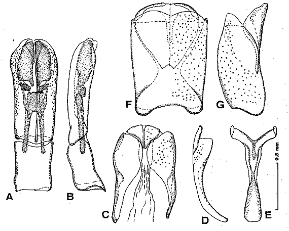
Fig. 21. Atholus philippinensis (Marseul). Male genitalia. A: Aedeagus, dorsal view. B: Ditto, lateral view. C: Ninth and 10th tergites, dorsal view. D: Ditto, lateral view. E: Ninth sternum (spicule), dorsal view. F: Eight tergite and sternum, dorsal view. G: Ditto, lateral view. [no. 9908, western Kalimantan, Indonesia]. (Ohara, 1999b).
MO-02-014 [Malayasia: Sapong, Sabah].